Joint pain can come from an injury, worn cartilage, inflammation, a tendon or ligament problem, referred nerve pain, or infection, and each cause calls for a different response. By the end, you will know which symptoms need urgent care, how clinicians investigate the source, what treatment usually comes first, and when surgery or coordinated rehabilitation becomes appropriate.
Key takeaways
- Seek emergency care for a hot, swollen joint with fever or severe movement loss.
- Orthopedic evaluation combines symptom history, movement tests, and targeted examination.
- X-rays show bone changes; MRI helps assess soft tissue and cartilage.
- Physiotherapy, medicines, injections, and activity changes often precede surgery.
When does joint pain need urgent care, and who should you see?
A hot, swollen joint with fever, severe pain, or restricted movement needs emergency assessment because septic arthritis can destroy a joint quickly. Emergency care also fits a visibly deformed joint, inability to bear weight, sudden loss of movement, numbness or weakness, or severe pain after a fall.
These symptoms make urgent orthopedic care necessary; do not wait for a routine appointment.
- Go to an emergency department after a major injury, especially with a deformed joint, suspected fracture, inability to bear weight, or a cold, pale, numb, or weak limb.
- Seek same-day assessment for a hot, swollen joint, fever, chills, or rapidly worsening pain. Clinicians may need to remove synovial fluid with a needle for cell-count testing and culture.
- Arrange prompt orthopedic review for persistent pain after an injury, locking, repeated giving-way, suspected tendon or ligament damage, or movement loss.
- Book primary-care care for gradual, stable pain without red flags, including suspected osteoarthritis. The clinician can examine you, manage initial treatment, and refer you.
- Ask for rheumatology review when several joints are swollen, morning stiffness lasts more than 30 minutes, symptoms recur, or gout or inflammatory arthritis is suspected.
- See a physiotherapist after serious injury and infection have been excluded, particularly for weakness, balance, gait, or movement limitations.
This joint pain triage is not based on an X-ray alone. An orthopedic surgeon evaluates injury, stability, strength, and function; a physiotherapist restores movement and gait; rheumatology investigates immune-driven inflammation.
How does an orthopedic evaluation identify the source of pain?
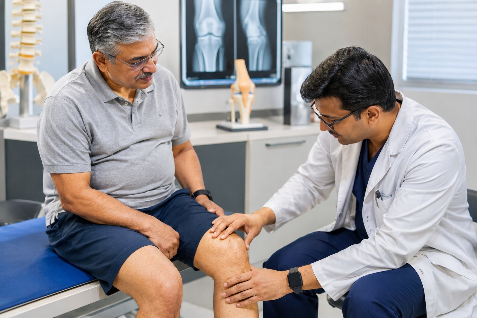
An orthopedic evaluation begins with a symptom history: when pain started, whether an injury occurred, where it spreads, and what worsens or relieves it. The clinician records swelling, redness, fever, morning stiffness, night pain, and activity limitations such as difficulty climbing stairs, gripping, or bearing weight.
Movement testing measures range and pain; strength assessment checks muscle function, while joint stability checks look for abnormal looseness.
- Acute injury: sudden pain after a fall, twist, or impact, often with bruising, swelling, deformity, or immediate loss of function.
- Osteoarthritis: gradual, activity-related pain with brief stiffness after rest; examination may show reduced range and creaking without major warmth.
- Inflammatory arthritis: prolonged morning stiffness, persistent swelling, warmth, fatigue, or several affected joints, prompting rheumatology assessment.
- Gout: abrupt, severe pain in a hot, red, swollen joint, commonly the big toe; joint-fluid testing can identify crystals.
- Tendon or ligament injury: pain during a specific resisted movement or stretch, with weakness or instability after overuse or trauma.
- Referred spine pain: back or neck symptoms with radiating pain, tingling, numbness, or weakness despite little swelling at the joint.
- Infection: a hot, swollen joint with severe restriction and fever is septic arthritis until assessed; urgent joint aspiration for synovial-fluid analysis and culture is central.
A visibly deformed joint, inability to bear weight, sudden loss of movement, numbness, weakness, or severe pain after a fall requires urgent care rather than a routine appointment.
Which imaging test is useful, and when can imaging mislead you?
An X-ray is the practical first choice for suspected fracture or arthritis, especially when pain follows an injury or weight-bearing is difficult. For uncomplicated chronic knee pain, weight-bearing radiographs show joint-space narrowing and bone changes more usefully than an immediate MRI.
| Test | Best use | What can mislead you |
|---|---|---|
| X-ray | Fracture and arthritis; weight-bearing radiographs for load-related pain | It cannot show most soft-tissue injuries, and visible wear may not match pain |
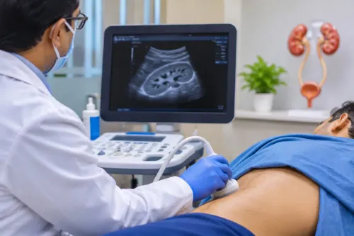
| Ultrasound | Joint fluid, tendon problems, and guided fluid aspiration | It depends on operator skill and cannot assess deep bone or every internal structure |
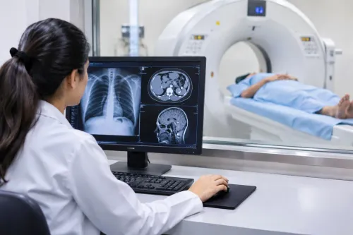
| MRI | Ligaments, cartilage, menisci, and marrow; suspected internal derangement or unexplained symptoms | It reveals abnormalities that may be painless and incidental |
Use ultrasound when swelling suggests joint fluid or a tendon problem, particularly when a clinician needs to guide aspiration. Reserve MRI for suspected ligament, cartilage, meniscus, or marrow disease, occult injury, or symptoms that remain unexplained after examination and initial radiographs.
Imaging does not measure pain, strength, stability, or the ability to climb stairs, walk, work, or bear weight. A scan can show arthritis in someone with little discomfort, while severe pain can occur without dramatic X-ray changes. The clinician must match the image with tenderness, movement range, weakness, instability, symptom timing, and functional limitation.
A hot, swollen joint with fever needs urgent assessment and joint-fluid testing; an MRI must not delay that evaluation.
What treatments are tried before joint surgery?
Care usually progresses from reducing joint stress and load to targeted pain relief, support, and active rehabilitation; surgery enters the discussion only when pain and disability persist despite an adequate trial.
1. Start with activity modification: reduce impact, kneeling, stairs, or lifting that triggers pain, while keeping tolerable movement. Weight management reduces load on weight-bearing joints, and pacing prevents overdoing activity on good days.
2. Choose medication based on the joint and your health. Topical NSAIDs are commonly preferred for knee osteoarthritis because they limit whole-body exposure. Oral NSAIDs require review of stomach, kidney, liver, and cardiovascular risks; use the lowest effective dose for the shortest practical period, often with stomach protection.
3. Add support when it improves function. A brace, cane, crutch, or walker can reduce painful loading and improve balance, but incorrect height or prolonged dependence can create new strain. A clinician or physiotherapist should fit the device.
4. Use injections selectively. Corticosteroid injections can provide short-term relief during a painful flare, allowing movement and therapy, but they do not repair worn cartilage.
5. Build capacity through exercise-based rehabilitation: range-of-motion work, strengthening, balance, gait practice, and home exercises. Measure progress by walking, stairs, sleep, and daily tasks—not an X-ray alone.
If these measures fail, reassess the diagnosis and function before discussing joint replacement or another procedure. Routine arthroscopic washout or debridement is not a lasting substitute for rehabilitation in degenerative knee osteoarthritis.
When does surgery or coordinated rehabilitation restore mobility more effectively?
Joint replacement becomes reasonable when pain, stiffness, and loss of function seriously restrict daily life despite an adequate trial of exercise, medication, injections, or support devices. Base the decision on symptoms and disability, not an X-ray alone; advanced wear without meaningful symptoms does not justify surgery.
Hip fracture surgery is urgent because delay increases medical complications and loss of independence. For an older adult admitted with a hip fracture, NICE recommends operating on the day of admission or the following day when the patient’s medical condition permits.
Recovery works better when movement begins early rather than waiting for complete pain relief. A coordinated rehabilitation plan may include:
- Early mobilization: sitting up, standing, and walking with the prescribed aid as soon as the surgeon and medical team allow.
- Progressive strengthening: graded exercises for the thighs, hips, trunk, and shoulder muscles, increasing resistance without provoking a damaging pain flare.
- Gait retraining: correcting stride, balance, weight-bearing, stair use, and safe handling of a walker or cane.
- Range-of-motion work and activity education: protecting the repair while gradually restoring movement and endurance.
- Home planning: arranging equipment, transport, bathroom safety, family support, and follow-up before discharge.
Physical and occupational therapists coordinate with orthopedic surgeons, nurses, pain or anesthesia teams, physicians managing other illnesses, and rehabilitation specialists. Hinjawadi SuperSpeciality Hospital 08048033666 can help coordinate orthopedic review and physiotherapy when medical conditions or home limitations make recovery more complex.
Related service
Orthopedic ₹500 Our Orthopedic Department provides comprehensive care for conditions affecting the bones, joints, muscles, ligaments, and spine. |
Frequently asked questions
When does joint pain need urgent care?
Seek emergency assessment for a hot, swollen joint with fever, severe pain, restricted movement, visible deformity, inability to bear weight, sudden movement loss, numbness, weakness, or severe pain after a fall.
How does an orthopedic evaluation find the cause of joint pain?
The evaluation reviews how the pain began, examines swelling and alignment, tests movement and strength, and uses targeted imaging or other tests when the findings require them.
Which imaging test is useful for joint pain?
X-rays are useful for fractures, alignment, and arthritis-related bone changes. MRI provides more detail about cartilage, ligaments, tendons, and other soft tissues.
When can imaging mislead you?
Imaging findings do not always explain pain. Age-related changes can appear in people without symptoms, so scan results must match the history and physical examination.
What treatments are tried before joint surgery?
Treatment can include activity changes, pain relief, physiotherapy, strengthening, weight management when relevant, supports, and injections selected for the diagnosed condition.