Early imaging can reveal a small nodule, hidden bleeding, a blocked vessel, or a tissue change before the problem produces obvious symptoms. You will learn when imaging is used for screening, when it is ordered because of a clinical concern, what each examination can uncover, and why an image alone does not settle the diagnosis.
Key takeaways
- Imaging can reveal structural changes before severe or obvious symptoms appear.
- Screening depends on age, risk factors, symptoms, and established clinical guidance.
- An abnormal scan needs clinical review; it does not prove a diagnosis.
- Doctors combine imaging with symptoms, examination, and test results before choosing care.
What makes imaging an early-detection tool?
Imaging supports early detection by showing structural changes before disease causes severe or obvious symptoms. A scan can reveal a small lung nodule, a breast mass too small to feel, silent kidney stones, fatty liver, vascular narrowing, or a bone lesion.
This makes radiology useful for finding asymptomatic disease, not only explaining illness after it progresses.
- Screening imaging tests people who have no symptoms but belong to a group with meaningful disease risk. A screening mammogram, low-dose CT for eligible high-risk smokers, or DXA bone-density scan looks for an abnormality before a complaint brings the person to a doctor.
- Diagnostic imaging investigates a specific concern. Symptoms, an examination finding, family history, or an abnormal blood test can prompt an ultrasound, CT, MRI, or X-ray to identify the cause, define its extent, or guide treatment.
- A screening result is not a diagnosis. A suspicious finding can require repeat imaging, contrast imaging, biopsy, or specialist review, while a normal scan cannot exclude every disease.
Early detection improves options only when the finding changes care. Incidental abnormalities can be harmless yet lead to surveillance scans or invasive tests, so doctors interpret the image alongside age, risk factors, symptoms, and the likely benefit of acting.
Which scans can reveal early changes?
A scan can expose a structural change before it produces obvious symptoms, but each method is strongest for a different target. The same finding can also be incidental, leading to repeat imaging or biopsy without proving disease.
| Method | Early conditions it can reveal | What it shows best |
|---|---|---|
| Mammography | Early breast cancer or suspicious breast abnormalities | Tiny masses, architectural distortion, and microcalcifications before a lump is palpable |
| Chest X-ray | Selected lung findings, pneumonia, fluid, or a large mass | Overall lung fields and heart size; it can miss small or hidden lesions |
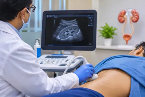
| Ultrasound | Gallstones, fatty liver, thyroid nodules, pregnancy findings, kidney problems, or vascular narrowing | Real-time soft tissue, fluid, blood flow, and fetal development without radiation |
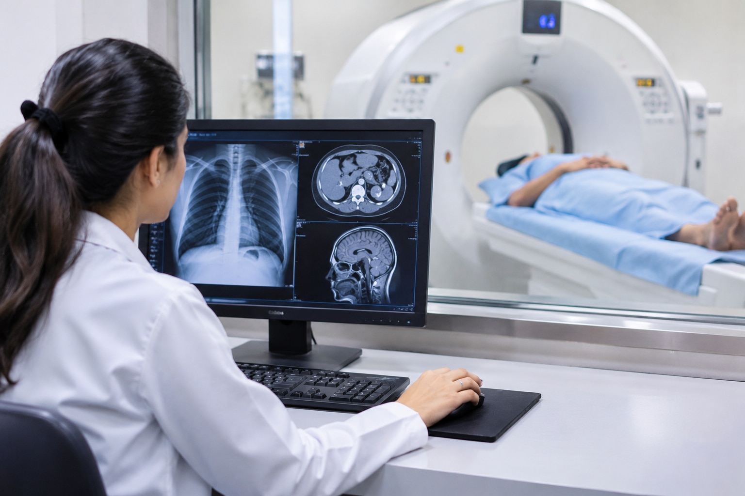
| CT scan | Small lung nodules, acute bleeding, early stroke complications, kidney stones, and complex anatomy | Rapid cross-sectional detail; CT angiography can show blocked vessels, while a normal early scan does not exclude every small stroke |
| MRI | Early changes in the brain, spine, joints, soft tissues, and selected tumors | Subtle tissue injury, cartilage, nerves, marrow, and tumor extent without ionising radiation |
| DXA | Low bone mineral density and osteoporosis risk | Bone density, not a guarantee that a fracture will or will not occur |
| PET-CT | Active cancer sites, spread, or recurrence | Increased glucose metabolism alongside anatomy; infection and inflammation can also appear bright |
Who should receive screening, and when?
Screening is for people without symptoms who meet an evidence-based age or risk threshold. The main situations are:
- Mammography: For average-risk women, mammography age 40 to 74 is recommended every other year. Dense breasts can reduce sensitivity and do not automatically justify ultrasound or MRI after a normal result.
- Low-dose CT lung cancer screening: Have an annual scan from ages 50 to 80 if you have a 20 pack-year smoking history and currently smoke or quit within 15 years. Stop after 15 smoke-free years or if illness prevents curative surgery.
- CT colonography: Average-risk adults aged 45 to 75 can choose this examination every five years. It requires bowel preparation, and a positive result requires colonoscopy for biopsy or polyp removal.
- Abdominal aortic aneurysm ultrasound: A one-time abdominal ultrasound is recommended for men aged 65 to 75 who have ever smoked. Other patients need individual risk assessment.
- DXA: Screen women aged 65 and older for osteoporosis; screen younger postmenopausal women when risk factors make a fracture risk assessment positive. Evidence is insufficient for routine screening of men.
A screening scan can trigger repeat imaging or invasive testing, so age, smoking exposure, family history, and health status matter.
How symptoms and test results trigger earlier imaging
Symptom-led imaging is diagnostic, not routine screening: the complaint, examination, risk profile, and urgency determine both timing and modality. A mild, persistent change may justify an outpatient scan; a sudden neurological deficit requires immediate imaging because treatment decisions depend on minutes.
- A new focal neurological sign or palpable lump prompts targeted imaging. Ultrasound can assess a superficial mass, thyroid, or vascular narrowing; mammography or MRI may follow when breast tissue or deeper structures need clearer assessment.
- Family history can move imaging earlier or change the test. A strong inherited cancer pattern, for example, may warrant specialist-directed MRI or another surveillance plan rather than waiting for symptoms.
- Abnormal laboratory results focus the search. Raised liver enzymes can lead to ultrasound showing fatty liver; blood in urine can lead to imaging that finds silent kidney stones or another urinary cause.
- Sudden weakness, speech trouble, severe headache, or loss of balance demands emergency assessment. For acute stroke, noncontrast CT rapidly checks for bleeding, CT angiography looks for a blocked large artery, and CT perfusion estimates threatened brain tissue. A normal early CT does not exclude a small infarct.
Early imaging can also reveal small lung nodules, but incidental findings often require comparison scans rather than immediate treatment.
What imaging can miss, and what an abnormality does not prove
A normal scan lowers the probability of disease; it does not exclude it. A lesion can be too small to resolve, appear after the scan, or lie outside what that test shows well. Dense breast tissue can hide a breast cancer on mammography, so persistent symptoms still need clinical review even after a normal result.
Repeating or changing the test also has trade-offs. CT adds a CT radiation dose, while MRI uses no ionizing radiation but requires MRI safety screening for certain implants or metal fragments. Gadolinium contrast is not needed for every MRI and requires extra consideration with severe kidney disease or a previous contrast reaction.
An abnormality is not a diagnosis. FDG-PET detects increased glucose use, but infection and inflammation can also appear bright, producing false-positive results. Incidental findings unrelated to your symptoms can trigger repeat scans, specialist appointments, or biopsy even when they are harmless.
Doctors judge the finding against its appearance, your age, risk factors, and whether acting on it will change care.
How doctors turn a scan into a care decision
A radiology report usually contains the clinical history, technique, contrast used, findings, impression, and follow-up recommendation. The findings describe what appears on the images; the impression ranks the most likely explanations and identifies urgent abnormalities. Comparison with prior scans can show whether a nodule, cyst, or lesion is new, growing, or stable.
The impression is not always the final diagnosis because imaging patterns overlap. Infection can resemble cancer, scar tissue can resemble active disease, and an incidental finding can be harmless despite appearing early. Doctors combine the report with symptoms, examination, laboratory results, age, and risk factors.
The next step depends on what remains uncertain:
- Comparison with prior scans may end the work-up when a finding is unchanged over an appropriate interval.
- Contrast imaging can clarify blood vessels, organ boundaries, inflammation, or a mass when a non-contrast scan is inconclusive.
- Biopsy samples tissue when cancer or another condition requires microscopic confirmation.
- Endoscopy may follow a bowel or stomach finding because it allows direct viewing and biopsy.
- Specialist referral follows when the finding needs surgery, oncology, pulmonology, gastroenterology, or another focused opinion.
At Hinjawadi SuperSpeciality Hospital 08048033666, ask which result changed the plan, what uncertainty remains, and why the proposed next test is safer or more useful than simply repeating the scan.
Related service
Radiology ₹1000 Radiology is a vital medical specialty that uses advanced imaging techniques—such as X-rays, CT scans, MRIs, and ultrasounds—to diagnose and treat... |
Frequently asked questions
What makes imaging useful for early detection?
Imaging shows structural changes that can appear before a condition causes severe or obvious symptoms, such as a small lung nodule or an early breast mass.
Which scans can reveal early health changes?
Depending on the suspected condition, doctors may use X-rays, ultrasound, mammography, CT, MRI, or other specialised imaging tests.
Who should receive screening, and when?
Screening decisions depend on factors such as age, personal and family history, known risks, and established screening guidance. Screening is not appropriate for every person or every scan.
How do symptoms and test results lead to earlier imaging?
Symptoms, examination findings, or abnormal blood and urine tests can give doctors a reason to order imaging before a condition becomes advanced.
What can imaging miss, and what does an abnormality prove?
Scans can miss small, early, or poorly visible changes. An abnormality is a finding, not a diagnosis; doctors may need comparison images, laboratory tests, follow-up imaging, or a biopsy.